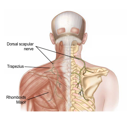
Upper Back Anatomy / Nerves Of The Chest And Upper Back Anatomy Of The Nerves Of The Chest And Upper Back Anatomy Medicine Com. The complexity of this region means that dysfunction can occur either due to injury or progressive pain and degeneration. It runs from the neck to the upper back. Evenly distribute weights from your upper body into the lower extremities. In the upper back region, the trapezius, rhomboid major, and levator scapulae muscles anchor the scapula and clavicle to the spines of several vertebrae and the occipital bone of the skull. It is like that for several reasons, all of which you can understand by looking at the anatomy of the thoracic spine.
The spine is made up of 33 individual bones called ve. Sometimes you feel the effects right away. They originate from the vertebrae and insert into the scapulae. Human anatomy · july 23, 2016. It is very stiff, and the thoracic spine has a limited range of motion.
Understanding Spine Anatomy Rojeh Melikian M D from rojehmelikianmd.com This muscle is located on the upper portion of the back anatomy, underneath the trapezius. Try the injurymap exercise app now. Human anatomy · july 23, 2016. It is very stiff, and the thoracic spine has a limited range of motion. Powerful muscles that move the head and arms attach to these bones as well. The upper back originates at the base of your neck, incorporates both shoulders and extends down to mid spine, including your ribs. The extrinsic (superficial) back muscles, which lie most superficially on the back. Sometimes you feel the effects right away.
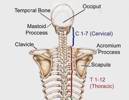
These include the cervical vertebrae in the neck, the thoracic vertebrae of the ribcage in the upper and middle back, the lumbar vertebrae in the lower back, and the vertebrae that are part of the pelvis.
In the upper back region, the trapezius, rhomboid major, and levator scapulae muscles anchor the scapula and clavicle to the spines of several vertebrae and the occipital bone of the skull. It contains many muscles and nerves but only has one bone, the femur, which is the longest and strongest bone in. The bones have a crystalline it, essentially, floats off of the back of the chest, as it is connected to the body primarily by muscle. 3d video tutorials and interactive modules on the anatomy of the vertebral. The lumbar and sacrum region make up the bone of the lower back anatomy. The rhomboid muscle is activated as you bring and squeeze your scapula or shoulder blades back and together. It is like that for several reasons, all of which you can understand by looking at the anatomy of the thoracic spine. Sometimes you feel the effects right away. Looking for a solution to your back pain problem? The main superficial muscles of the back are the following: In order to understand why upper back pain occurs, it's helpful to know the basic anatomy of the spine. The complexity of this region means that dysfunction can occur either due to injury or progressive pain and degeneration. The trapezius and latissimus dorsi muscles connect the upper limb to the vertebral column.
The rhomboid muscle is activated as you bring and squeeze your scapula or shoulder blades back and together. Balance the weight of your head on top of your spine. The spine is made up of 33 individual bones called ve. Upper_back_anatomy_chart 6/23 upper back anatomy chart emotional pain for good. Back anatomy the back is the body region between the neck and the gluteal regions.
What Are The Bones Called In Your Neck Shoulder Area And Upper Back Socratic from usercontent2.hubstatic.com The spine is made up of 33 individual bones called ve. The basic anatomy of your upper back by lindsey mcfadden as you're doing your regular upper back stretching exercises , you're probably wondering about the components of your upper back and why it happens to be the most stable part of your spine. These include the cervical vertebrae in the neck, the thoracic vertebrae of the ribcage in the upper and middle back, the lumbar vertebrae in the lower back, and the vertebrae that are part of the pelvis. 3d video tutorials and interactive modules on the anatomy of the vertebral. In the upper back region the trapezius rhomboid major and levator scapulae muscles anchor the scapula and clavicle to the spines of several vertebrae and the occipital bone of the skull. There is a set of muscles in the upper back (called the thoracic area) called the spinalis thoracis. It is like that for several reasons, all of which you can understand by looking at the anatomy of the thoracic spine. The bones of the chest and upper back combine to form the strong, protective rib cage around the vital thoracic organs such as the heart and lungs.
The lumbar and sacrum region make up the bone of the lower back anatomy.
The bones of the chest and upper back combine to form the strong, protective rib cage around the vital thoracic organs such as the heart and lungs. Your lower back (lumbar spine) is the anatomic region between your lowest rib and the upper part of the buttock. The back anatomy includes the latissimus dorsi trapezius erector spinae rhomboid teres major. The cervical spine is the top part of the spine. Upper_back_anatomy_chart 6/23 upper back anatomy chart emotional pain for good. In the upper back region the trapezius rhomboid major and levator scapulae muscles anchor the scapula and clavicle to the spines of several vertebrae and the occipital bone of the skull. In order to understand why upper back pain occurs, it's helpful to know the basic anatomy of the spine. It contains many muscles and nerves but only has one bone, the femur, which is the longest and strongest bone in. The rhomboid muscle is activated as you bring and squeeze your scapula or shoulder blades back and together. Extending from the base of the skull into the pelvis, it consists of 33 stacked bones known as vertebrae. License image the deltoid, teres major, teres minor, infraspinatus, supraspinatus (not shown) and subscapularis muscles (not shown) all extend from the scapula to the humerus and act on the shoulder joint. They originate from the vertebrae and insert into the scapulae. 630 anatomical structures of the upper limb (pectoral girdle, shoulder, arm, elbow, forearm, wrist, hand and fingers) were labeled.
The back functions are many, such as to house and protect the spinal cord, hold the body and head upright, and adjust the movements of the upper and lower limbs. It runs from the neck to the upper back. Both the deltoid and the trapezius are firmly attached to … Powerful muscles that move the head and arms attach to these bones as well. Extending from the base of the skull into the pelvis, it consists of 33 stacked bones known as vertebrae.
Shoulder Shoulder Blade Upper Back Pain And Or Pain Radiating Down The Back Of The Arm Could It Be Dns Solstice Physical Therapy from images.squarespace-cdn.com 3d video tutorials and interactive modules on the anatomy of the vertebral. See human back anatomy stock video clips. Back muscles anatomy here include the trapezius, latissimus dorsi, rhomboid and levator scapulae. The cervical spine is the top part of the spine. It contains many muscles and nerves but only has one bone, the femur, which is the longest and strongest bone in. It is like that for several reasons, all of which you can understand by looking at the anatomy of the thoracic spine. Anatomy of the back organs. Evenly distribute weights from your upper body into the lower extremities.
The cervical spine protects the nerves connecting to.
License image the deltoid, teres major, teres minor, infraspinatus, supraspinatus (not shown) and subscapularis muscles (not shown) all extend from the scapula to the humerus and act on the shoulder joint. All these muscles are therefore associated with movements of the upper limb. Balance the weight of your head on top of your spine. This muscle is located on the upper portion of the back anatomy, underneath the trapezius. 3d video tutorials and interactive modules on the anatomy of the vertebral. The spine is made up of 33 individual bones called ve. Before giving our recommendations for upper back exercises, it's important to first go over the anatomy of the back musculature. It contains many muscles and nerves but only has one bone, the femur, which is the longest and strongest bone in. The bones of the chest and upper back combine to form the strong, protective rib cage around the vital thoracic organs such as the heart and lungs. 1 your spine in this region has a natural inward curve. Human body anatomy female female anatomy muscle shoulder blade pain anatomy back muscles bones man female anatomy body muscles in a body female anatomy muscole shoulder concept muscular sysyem. They originate from the vertebrae and insert into the scapulae. There is a set of muscles in the upper back (called the thoracic area) called the spinalis thoracis.